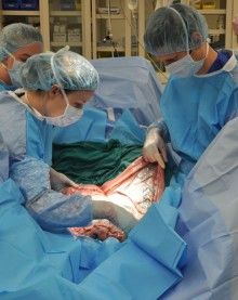
CALEC Surgery: New Hope for Corneal Damage Treatment
In the realm of ocular regenerative medicine, CALEC surgery stands out as a groundbreaking advancement in treating corneal damage. This innovative procedure utilizes stem cell therapy, involving the cultivation of limbal epithelial cells harvested from a healthy eye, to repair and restore severely damaged corneal surfaces. Clinically tested with promising results, CALEC surgery has shown effectiveness in alleviating chronic pain and restoring vision in patients whose conditions were previously considered untreatable. The meticulous process of transplanting these cultivated cells reveals new hope for individuals affected by severe eye injuries and related visual impairments. As research progresses, CALEC surgery represents a significant leap forward in eye injury therapy, potentially transforming lives through enhanced ocular health and recovery.
Known as cultivated autologous limbal epithelial cell therapy, CALEC surgery is revolutionizing treatments for those suffering from corneal injuries. By harnessing the healing power of limbal stem cells, this technique enables the reconstruction of damaged ocular surfaces, offering a novel avenue for recovery in patients facing dire vision challenges. As the field of regenerative ophthalmology progresses, therapies like CALEC are poised to carve a new path for addressing the complexities of corneal damage, with research affirming its safety and efficacy. Not only does this method enhance the prospects for successful vision restoration, it also emphasizes the growing importance of stem cell applications in modern eye care. This pioneering approach fosters hope for a future where even the most severe ocular conditions may be effectively managed.
Understanding CALEC Surgery and Its Significance
CALEC, or cultivated autologous limbal epithelial cells, surgery represents a groundbreaking approach in the realm of ocular regenerative medicine. This innovative technique involves harvesting stem cells from a healthy eye to create a graft that can be transplanted into a damaged cornea, thereby restoring its surface. The significance of CALEC surgery lies not only in its potential to treat previously untreatable corneal damage but also in its ability to alleviate chronic pain and improve visual acuity for patients suffering from limbal stem cell deficiency.
The success of CALEC surgery is underscored by clinical trials conducted at Mass Eye and Ear, where a remarkable success rate was observed in restoring corneal surfaces. Patients who participated in this trial experienced a significant increase in their quality of life, providing hope for those who have long endured the debilitating effects of corneal injuries. As more data emerges, CALEC surgery continues to pave the way for advancements in stem cell therapy and eye injury therapy, reflecting a new era in ophthalmology.
The Role of Limbal Epithelial Cells in Ocular Health
Limbal epithelial cells are essential for maintaining the integrity and smoothness of the corneal surface. They reside at the outer edge of the cornea, the limbus, and play a critical role in responding to injuries and maintaining corneal clarity. When the eye sustains an injury, such as from chemical burns or infections, these stem cells can be depleted, leading to limbal stem cell deficiency. This condition severely impacts vision and can lead to chronic discomfort for affected individuals.
Research has shown that replenishing these limbal epithelial cells through methods such as CALEC surgery can reverse corneal damage and restore visual function. By utilizing advanced techniques in ocular regenerative medicine, the deficit of stem cells can be effectively addressed, offering a promising solution to patients with debilitating ocular injuries. This innovative approach emphasizes the importance of preserving and regenerating these vital cells to ensure long-term eye health.
Key Findings from Recent Clinical Trials on CALEC
Clinical trials conducted to evaluate the efficacy of CALEC surgery revealed compelling results, emphasizing its safety and effectiveness. In a study involving 14 participants, over 90 percent of patients experienced significant restoration of their corneal surfaces following the transplantation of the graft. These encouraging results demonstrate the potential of CALEC as a viable treatment strategy for individuals who suffer from severe corneal damage that was once deemed irreparable.
Additionally, the trials highlighted the ongoing need for larger studies to validate these findings further. Early results indicated that nearly half of the participants achieved complete restoration of their cornea within three months, and this success rate improved significantly over 18 months. Such promising outcomes not only advocate for CALEC surgery but also inspire further research into stem cell therapy in the context of ocular disorders.
The Process of Harvesting Stem Cells for CALEC
The procedure for harvesting limbal epithelial cells for CALEC surgery involves a precise method that balances safety and efficacy. By performing a biopsy on a healthy eye, surgeons can extract the necessary stem cells and subsequently expand these cells in a controlled environment to develop a robust cellular graft. This process, which takes approximately two to three weeks, ensures that the transplanted tissue meets the rigorous quality standards imperative for human use.
Once the graft is prepared, it is meticulously transplanted into the damaged cornea, where it integrates and begins to restore the eye’s surface. This innovative manufacturing process underscores the potential of ocular regenerative medicine, offering new hope to individuals suffering from debilitating eye conditions. As technology advances, the efficiency and accessibility of this process are likely to improve, further enhancing the prospects for patients with corneal damage.
Challenges and Limitations of CALEC Surgery
Despite its promising outcomes, CALEC surgery presents certain challenges that researchers must address before it can become widely available. One of the primary limitations is the requirement for patients to have only one healthy eye from which to harvest stem cells. This restriction poses a significant barrier for individuals with bilateral corneal damage, making it crucial for researchers to explore alternative sources for stem cells.
Efforts are underway to develop an allogeneic manufacturing process that would enable the use of limbal stem cells from donor eyes. This advancement could vastly expand the treatment’s applicability and provide opportunities for patients with damage to both eyes. As ongoing studies evolve, overcoming these limitations will be essential for maximizing the therapeutic potential of CALEC surgery and ultimately improving patient outcomes.
Future Directions in Limbal Stem Cell Research
The future of limbal stem cell research, particularly in the context of CALEC surgery, is brimming with potential. Ongoing clinical trials aim to involve larger patient populations and longer follow-up periods to gather comprehensive data regarding the surgery’s effectiveness. This increased scope not only enhances the reliability of the findings but also provides a foundation for potential FDA approval that could make CALEC widely accessible.
Moreover, innovative research in ocular regenerative medicine could lead to the discovery of new therapies and methodologies that complement CALEC. As scientists and medical professionals continue to collaborate and develop cutting-edge techniques, the landscape of eye injury therapy is poised for dramatic transformation, heralding improved treatments for those suffering from acute and chronic ocular conditions.
Understanding Ocular Regenerative Medicine
Ocular regenerative medicine is a rapidly evolving field dedicated to restoring vision and improving ocular health through innovative therapeutic techniques. Within this domain, stem cell therapy has emerged as a beacon of hope for individuals suffering from various types of eye injuries, including those resulting in corneal damage. By utilizing the body’s own cells, treatments like CALEC surgery underline the shift from traditional approaches to more regenerative strategies, paving the way for personalized and effective care.
This branch of medicine encompasses a wide array of techniques aimed at repairing or replacing damaged tissues within the eye. As research continues to unveil the complexities of limbal stem cells and their roles in maintaining corneal integrity, we can anticipate advancements that not only enhance existing treatments but also provide new avenues for addressing conditions previously viewed as untreatable. The future of ocular regenerative medicine encourages us to rethink our approach to eye health and injury.
The Importance of Collaboration in Ocular Research
Successful advancements in ocular research, particularly in the development of CALEC surgery, highlight the importance of collaboration among various institutions and disciplines. Research teams, such as those at Mass Eye and Ear, Dana-Farber, and Boston Children’s Hospital, work synergistically to bring innovative therapies to fruition. This collaborative spirit fosters an environment where shared knowledge and resources can lead to breakthroughs in treatment methods for eye health.
Moreover, partners across clinical and research settings can expedite the translation of laboratory discoveries into clinical applications, ensuring that promising therapies like CALEC surgery reach the patients who need them most. Continued collaboration among ophthalmologists, researchers, and regulatory bodies will be paramount in overcoming barriers and ultimately enhancing patient care through exciting new treatments.
The Future of Stem Cell Therapy in Ophthalmology
As we look towards the future, stem cell therapy’s role in ophthalmology is anticipated to expand dramatically. With successful trials demonstrating the efficacy of CALEC surgeries, the field is set to explore new indications for stem cell applications in various eye disorders. Increased research funding and focus from institutions dedicated to eye health underscore the growing recognition of stem cell therapy as a viable treatment option.
Innovations may not only improve the current methodologies but also introduce novel approaches that could address the complexities of ocular diseases. As researchers continue to decipher the pathways and mechanisms underlying ocular injury and regeneration, the hope is to promote healing and restoration for an ever-increasing patient population. The journey towards refining and popularizing stem cell therapies in ophthalmology is just beginning.
Frequently Asked Questions
What is CALEC surgery and how does it relate to stem cell therapy?
CALEC surgery, or Cultivated Autologous Limbal Epithelial Cell surgery, is a revolutionary procedure utilizing stem cell therapy to treat corneal damage. It involves harvesting healthy limbal epithelial cells from a patient’s unaffected eye, expanding these cells into a tissue graft, and then surgically transplanting this graft into the damaged cornea. This technique is particularly beneficial for individuals with severe corneal injuries, allowing restoration of the eye’s surface that was previously considered untreatable.
How effective is CALEC surgery for treating corneal damage?
Clinical trials have shown that CALEC surgery is over 90% effective in restoring the cornea’s surface. In a study following 14 patients for 18 months, complete restoration of the cornea was achieved in 50% of participants at three months, increasing to approximately 79% and 77% at twelve and eighteen months, respectively. This demonstrates significant potential for CALEC surgery in ocular regenerative medicine.
What are limbal epithelial cells and their role in CALEC surgery?
Limbal epithelial cells are specialized cells located at the cornea’s outer edge, responsible for maintaining a healthy corneal surface. In CALEC surgery, these cells are harvested from a healthy eye and cultivated to create a graft that can be transplanted into a damaged cornea, thus aiding recovery and restoring vision in those suffering from corneal damage.
Who is a candidate for CALEC surgery?
Candidates for CALEC surgery typically include individuals with significant corneal damage who have suffered from conditions like chemical burns, infections, or trauma that led to limbal stem cell deficiency. Notably, patients should have only one affected eye to allow for the necessary biopsy to collect limbal epithelial cells from the healthy eye.
Is CALEC surgery currently available, and what does the future hold?
As of now, CALEC surgery remains experimental and is not widely available in U.S. hospitals. Future studies aim to involve larger patient groups and further extend the findings of initial trials, with the hope of gaining federal approval to expand access to this innovative stem cell therapy for corneal damage.
What safety profile does CALEC surgery have according to clinical trials?
The clinical trials for CALEC surgery have reported a high safety profile, indicating minimal adverse events. Most participants experienced only minor complications, with no serious side effects documented in donor or recipient eyes. This encourages further exploration of CALEC as a viable solution for treating corneal injuries.
How does CALEC surgery contribute to the field of ocular regenerative medicine?
CALEC surgery represents a significant advancement in ocular regenerative medicine by providing a novel treatment option for patients with previously untreatable corneal damage. Through stem cell therapy, it offers hope for restoring vision and reducing pain in patients who would otherwise have limited options, marking a substantial step forward in eye injury therapy.
What are the next steps for CALEC surgery research?
Future research on CALEC surgery will focus on conducting larger clinical trials across multiple centers, longer follow-up periods, and establishing randomized control designs to validate the efficacy and safety of the procedure. These steps are crucial for advancing the treatment toward FDA approval and providing access to a broader patient demographic.
| Key Point | Details |
|---|---|
| First CALEC Surgery | Performed by Ula Jurkunas at Mass Eye and Ear, signaling a breakthrough in corneal injury treatment. |
| Clinical Trial Overview | Involved 14 patients, demonstrating safety and efficacy of CALEC in restoring corneal surfaces. |
| Procedure Details | Stem cells harvested from a healthy eye are expanded into grafts and transplanted into the damaged eye. |
| Effectiveness | Over 90% effective in restoring cornea, with significant improvements in visual acuity over time. |
| Safety Profile | High safety profile with minor adverse events; no serious complications reported. |
| Future Aspirations | Aim to develop an allogeneic process for patients with damage to both eyes. |
| Next Steps | Future studies will involve larger populations, longer follow-ups, and randomized designs. |
Summary
CALEC surgery represents a groundbreaking advancement in the treatment of corneal damage that was previously deemed untreatable. The positive outcomes of the initial clinical trial conducted at Mass Eye and Ear highlight its potential to significantly restore vision and improve quality of life for affected patients. As the research progresses toward FDA approval, there is hope for broader accessibility of this innovative stem cell therapy across the United States.

